Arterial Duplex Study

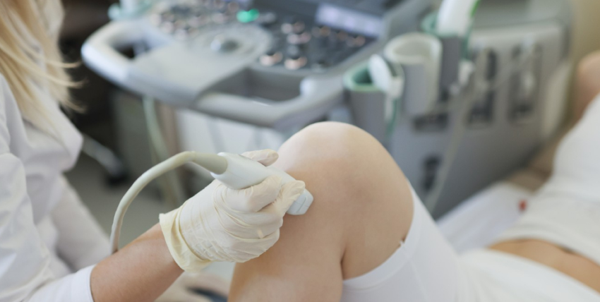
An Arterial Duplex Study is also known as LEAI (lower extremity arterial imaging). This test will check the arterial circulation in the legs. This non-invasive test does not use needles, drugs, or dyes. The test will be performed by a highly-skilled ultrasound technologist.
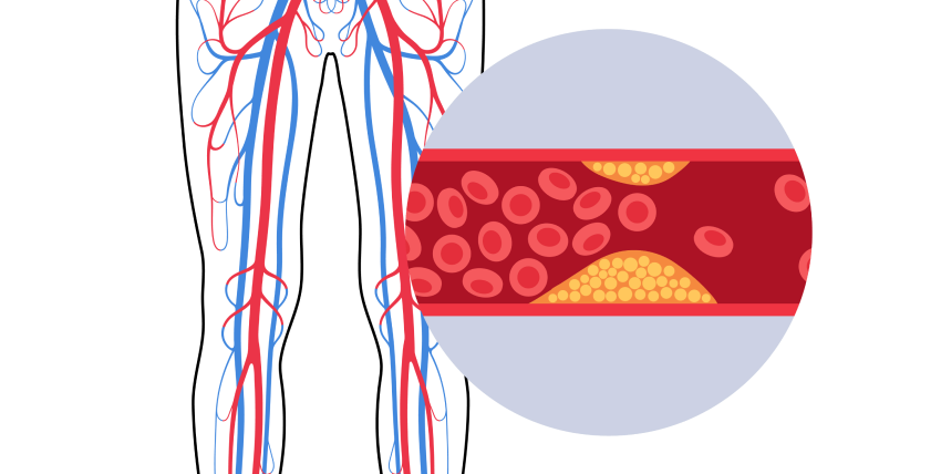
A small amount of gel is applied to the skin over the area to be studied. A handheld transducer is used to make the pictures. The pictures will show the walls of the arteries, blood flow through the arteries, and any plaque formation in the arteries. The velocity of the blood flow will be measured. This will pinpoint the level and severity of the disease if any.
Your doctor may order additional testing if this test is abnormal. This test always includes an ABI (ankle-brachial index). Blood pressures are taken in both arms and ankles. The pressures are compared and calculations performed to estimate the severity of the disease.

Adenosine Myocardial Perfusion Imaging Test

An Adenosine Myocardial Perfusion Imaging test uses a radioactive imaging agent (not a dye) to produce pictures of the heart muscle. The imaging agent gives off a small amount of radiation which can be seen with a special camera. The amount of radiation exposure during this test is very small and poses no health risk.
The test will help your doctor determine if there are areas of your heart that do not receive enough blood supply due to coronary artery disease. Areas of your heart that may have been damaged from a previous heart attack may also be seen.

Prior to the test, an IV will be started. During the rest portion of the test, you will receive an injection of the radioactive imaging agent, and pictures will be taken of your heart. For the stress portion of the test, a medication (Adenosine) will be given through the IV to simulate exercise by dilating the blood vessels of the heart. During the infusion, you will be continuously monitored for blood pressure, heart rate, rhythm, and ECG changes.

You will be given an injection of the radioactive imaging agent during the infusion. Following the Adenosine infusion, pictures will be taken to determine blood flow to the heart muscle. The test will help your doctor determine if there are areas of your heart that do not receive enough blood supply due to coronary artery disease. Areas of your heart that may have been damaged from a previous heart attack may also be seen.

Cardioversion

A cardioversion is done to change or “convert” an irregular heartbeat to a normal regular rhythm. The Heart The heart is a muscular organ about the size of a closed fist. It pumps oxygen-rich blood to the body.A cardioversion is done to change or “convert” an irregular heartbeat to a normal regular rhythm. The Heart The heart is a muscular organ about the size of a closed fist.
For the heart to do its work, it needs a “spark plug” or electrical impulse or signal from special tissue (sinus node) in the heart. The electrical impulse causes the heart muscle to contract. The hearts electrical system and muscle work together to effectively pump blood. Usually the heart beats regularly, but sometimes the heart beats irregularly. An irregular heart beat can occur after a heart attack or heart surgery. It may also occur with heart valve disease or other diseases not related to the heart. One cause of an irregular heart beat is atrial fibrillation. Some people with atrial fibrillation have no symptoms. Others may feel palpitations, shortness of breath, weakness or fatigue during episodes of atrial fibrillation. Your physician may first use medicines to convert your atrial fibrillation to a regular rhythm. If medicines do not work, an electrical cardioversion may be needed. This procedure uses small amounts of electrical current given through patches or paddles placed on the chest. The electrical current is used to restore your heart to a normal regular rhythm. When you are completely asleep, the physician will administer a very brief current of electricity. This electrical stimulation is intended to stop the irregular heartbeat and replace it with a normal heart rhythm. This is called converting the rhythm. The physician will administer the electrical current one to three times. Most people will convert to a normal rhythm with only one shock. If you do not convert after three attempts, the physician will not attempt any further electrical stimulation. The entire process lasts only a few minutes. The sedative medication will take from 10 minutes to two hours to wear off depending on the medication used and your individual response. The nurse will keep you in the room until you are completely awake and are able to sit up and talk. The nurse will make any follow-up appointments at this time and review any medication changes with you and your family members. On rare occasions the physician will administer special medications that assist in converting an irregular heart rhythm. If this occurs you may need to stay longer and be moved to another area that allows close monitoring of your heart rhythm for a few hours. This is a precaution that is used whenever this medication is administered.

Carotid U/S

Carotid ultrasonography (carotid U/S) is an ultrasound of the carotid arteries. Carotids are the arteries in your neck that supply blood to your brain. A small transducer, similar to the one used for listening with a stethoscope, is placed on the patient’s neck next to their windpipe (trachea). 
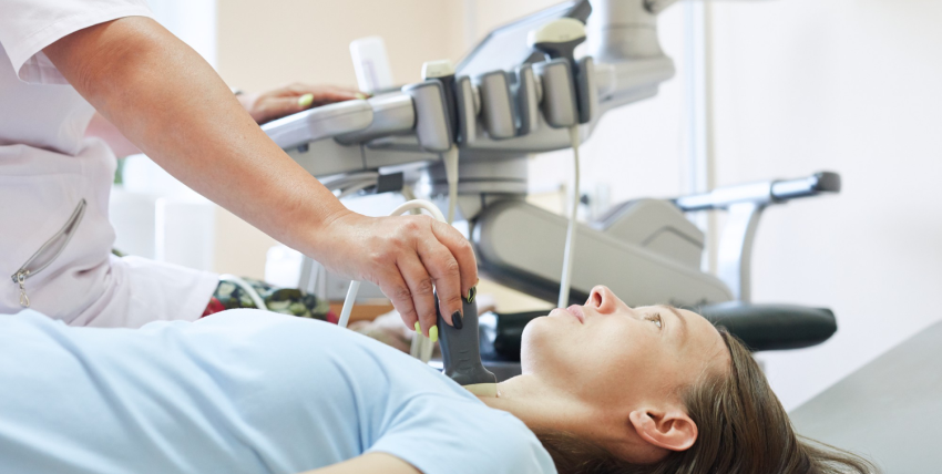
When it’s pressed firmly into place, the transducer emits sound waves that bounce back and create echoes when they hit objects – in this case, different types of tissue in the carotid artery.Carotid ultrasounds are most often done in order to look at plaque build-up inside of blood vessels (“carotid intima-media thickness”), but they can also be done to look for atherosclerotic plaques, blood clots, and narrowing of the carotid arteries. Carotid ultrasounds are usually done on people who have risk factors for or a history of stroke or transient ischemic attack (TIA).
Carotids ultrasound images may be interpreted by a sonographer and reported by an ultrasonographer or vascular technologist. Carotids U/S reports will often include measurements such as the thickness of the intima-media complex (IMT), which is used to assess plaque build-up. Carotids ultrasounds also provide information about any stenosis in the carotid artery, which can lead to decreased blood flow. Carotids U/S is also referred to as Carotids duplex U/S. Carotid ultrasounds are sometimes done in conjunction with carotid duplex Doppler ultrasounds, another type of ultrasound that provides information about blood flow in the arteries. Carotids duplex Doppler ultrasounds are most often used to look for carotid artery stenosis or occlusions. Ultrasound is a painless procedure that takes very little time.
Carotid percussions can be performed at the bedside by compressing the neck over the artery and producing a thudding sound when it is released. This technique has been replaced by high-frequency ultrasound because it is more sensitive but less specific in detecting vascular abnormalities . Carotid auscultation has been shown to have a specificity of less than 50% for identifying vascular pathology. Carotids ultrasounds may be used to look at the following: Carotids arch – Carotids bifurcation – Carotid body – Carotids endarterectomy – Carotids “halo sign” – Carotid intima-media thickness (IMT) – Carotid lumen stenosis/obstruction – Carotids plaque build-up – Carotid plaques, calcified plaques, and complex composite plaques. Carotid Artery Ultrasound Images. Carotid artery ultrasound image showing noncompliant left carotid wall.

Cardiac Catherization

A Cardiac Catheterization allows a physician to examine a patient’s heart for pumping function, check for blockage in the arteries bringing blood to the heart, and measure pressures within the heart. Patients are usually given a mild sedative prior to the procedure. This will help you feel more relaxed and maybe a bit sleepy.This test is done in a room with special cameras to take x-ray pictures of your heart. 
The nurse or technician will cleanse and shave the area and cover you with sterile drapes. The doctor will then numb the area and insert a small hollow tube or sheath into the artery in your groin (or arm). The physician can then thread special catheters (long, thin plastic tubes) up to the heart.This test is done in a room with special cameras to take x-ray pictures of your heart. Most cardiac catheterizations are done through a large artery in the groin, However Dr. De is known for being one of the few who prefer the radial approach, done through the wrist.


Through these specially shaped catheters, the physician can inject x-ray contrast dye and take x-ray pictures of the arteries that supply blood to the heart muscle and the main pumping chamber of the heart. Some people may notice a mild discomfort or “hot flash” following the dye injection which will last only a few seconds.


During the catheterization, the physician can view the x-ray images on special TV screens. The entire procedure can take as little as 20 minutes or sometimes more than an hour, depending on how many and what type of measurements are taken. After the physician has completed the procedure, the sheaths are removed from the groin (or arm) and a nurse or cath lab technician will hold pressure on the site to allow the small hole in the artery to close. A special dressing, pressure device or sandbag may be placed on the site at this time.


You will be asked to lie flat and still for several hours after the procedure to allow the hole in the artery to seal. Some physicians use a type of vascular closure device to seal the puncture in the artery. You should follow your physician’s specific instructions about activity following the use of one of these devices.

Most patients will go home several hours after the procedure, but because you have been given medication to relax you, please plan to have someone drive you home.
Coronary Angioplasty & Stents

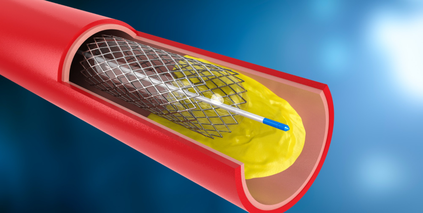
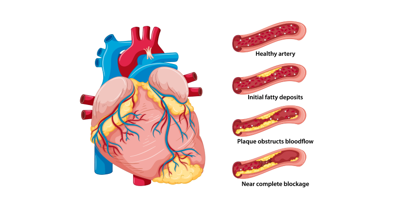
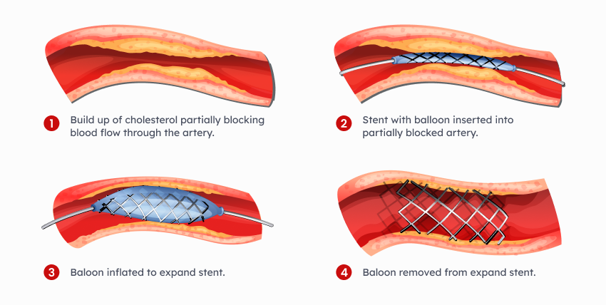
A Coronary Angioplasty (PTCA) is a procedure in which a physician utilizes a small balloon catheter (flexible plastic tube) to open up a blockage in an artery that supplies blood to the heart muscle. Frequently a small, metal, spring-like device called a stent is also implanted into the coronary artery to reduce the chance that the blockage will recur. Coronary Angioplasty/Stent can relieve blockages to allow a person to be active without chest discomfort and prevent a heart attack. Patients are usually given a mild sedative prior to the procedure. This will help you feel more relaxed and maybe a bit sleepy.
Through these specially shaped catheters, the physician can thread a wire across the blockage in the artery. Then, a thin expandable balloon is placed over the wire and inflated to open up the blockage. Several balloons of increasing size may be used to reduce the blockage. During a stent procedure, the stent, which is tightly mounted on a balloon, is placed across the blockage and the balloon is inflated opening the stent. Several other devices may be used during the procedure. Your physician will discuss these options with you.
Some people may notice mild chest discomfort while the balloon is inflated. You may be given a medication through your IV for several hours to reduce the chances of blood clots forming. The entire procedure can take as little as 30 minutes or sometimes more than an hour. After the procedure, most patients will be transferred to a post-procedure care area where the sheaths are removed from the groin when the blood thinners have worn off. You will be asked to lie flat and still for several hours after the procedure to allow the hole in the artery to seal. Some physicians use a type of vascular closure device to seal the puncture in the artery. You should follow your physician’s specific instructions about activity following the use of one of these devices. Most patients will stay in the hospital overnight.

Implantable Cardiac Monitor (Loop Recorder)

Your cardiologist may recommend surgically implanting a wireless cardiac monitor called a loop recorder, which continuously records your heart’s rhythm for up to three years. Smaller than the size of an AAA battery, this device is surgically inserted beneath the skin of the upper chest to record the heart’s electrical activity, much like an electrocardiogram, or EKG.

People who have had unexplained fainting spells or heart palpitations that can’t be detected by short term heart rhythm recording devices—such as Holter monitors—may be candidates for this device. Doctors may also recommend an implantable loop recorder for people with atrial fibrillation, which causes a rapid and irregular heartbeat. In addition, this device is used in people who have had a stroke for which a cause has not been determined.


Your doctor makes a small incision in the upper chest and inserts the device just under the skin. This is a brief, outpatient procedure done in our office, you can return to your usual activities the next day. Your doctor may limit your activity until the incision heals.


Cardiac loop recorders typically monitor the heart’s electrical activity for up to 36 months before replacement is required. The device is removed through the original incision site through a short surgery that is similar to the implantation procedure.

Mobile Cardiac Telemetry

Mobile Cardiac Telemetry is when a monitor is placed on a patient to transmit the heartbeat patterns remotely and in real-time. Mobile Cardiac Telemetry (MCT) is the cutting-edge of cardiac monitoring. MCT devices are small portable monitors that send a constant feed of data showing the ECG (Electrocardiography) readout of the patient in real-time.
Electrocardiography is the process of recording the electrical activity of the heart over a period of time using electrodes placed over the skin. These electrodes detect the tiny electrical changes on the skin that arise from the heart muscle’s electrophysiologic pattern of depolarizing and repolarizing during each heartbeat.

Peripheral Vascular Interventions

What is Peripheral Vascular Intervention?

Peripheral vascular intervention is a minimally invasive outpatient (same day) procedure used to treat peripheral artery disease, which causes plaque buildup in the arteries leading to the intestines, head, arms, and most commonly the legs. Plaque buildup is also called atherosclerosis.

The goal of peripheral vascular intervention is to restore the flow of blood to your lower extremities, eliminating pain, numbness, or need for amputation. Our specialists typically employ two different methods – angioplasty and atherectomy.


Angioplasty & Stents
Angioplasty is a minimally invasive intervention method that uses a balloon-tipped catheter to dilate your artery. Once inserted, the balloon is inflated at the blockage site to compress the plaque against the wall of the artery, increasing the flow of blood. In order to prevent the artery from becoming blocked again long-term, a small tube called a stent is sometimes put in place.


Atherectomy
Atherectomy is a non-surgical intervention that removes plaque using a catheter with either a sharp blade or a small drill on its tip. The plaque is either captured by the end of a catheter or is broken up and safely released into the bloodstream.

Positron Emission Tomography (PET)

What is a cardiac PET scan?

A PET scan of the heart is a noninvasive nuclear imaging test. It uses radioactive tracers (called radionuclides) to produce pictures of your heart. Doctors use cardiac PET scans to diagnose coronary artery disease (CAD) and damage due to a heart attack. PET scans can show healthy and damaged heart muscle. Doctors also use PET scans to help find out if you will benefit from a percutaneous coronary intervention (PCI) such as angioplasty and stenting, coronary artery bypass surgery (CABG) or another procedure.

Quick Facts

  • PET scans use radioactive material called tracers. Tracers mix with your blood and are taken up by your heart muscle.
  • A special “gamma” detector that circles the chest picks up signals from the tracer. A computer converts the signals into pictures of your heart at work.
  • A PET scan shows if your heart is getting enough blood or if blood flow is reduced because of narrowed arteries. It also shows dead cells (scars) from a prior heart attack.
  • A PET scan can help in determining if you’ll benefit from a cardiac procedure (PCI) or surgery to restore blood flow. The tracers used for PET scans can help identify injured but still living (viable) heart muscle that might be saved if blood flow is restored.
  • Why do people have cardiac PET scans?

    A PET scan is a very accurate way to diagnose coronary artery disease and detect areas of low blood flow in the heart. PET can also identify dead tissue and injured tissue that’s still living and functioning. If the tissue is viable, you may benefit from a PCI or coronary artery bypass surgery.

    How does a PET scan work?

    A radioactive tracer is injected into your bloodstream. The tracers used for PET are mostly natural body compounds such as glucose, water or ammonia, which are labeled or “tagged” with a small amount of radioactive material. Inside your body the radioactive tracer produces a type of energy called a gamma ray. Gamma rays are detected by a gamma detector and are used to produce a series of clear images of your heart. Images of thin slices made all the way through the heart can be produced from all different directions and angles.
    Computer graphics can be used to create a 3-dimensional image of your heart from the thin-slice images. Your doctor will be able to tell if your heart muscle is functioning or (if the heart muscle isn’t working) if your heart muscle is still alive by how well it takes up and uses the different tracers. The doctor will determine this by examining the images to find where the tracer is. Viable heart tissue will take in more of the tracer than tissue that’s no longer viable. Different colors or degrees of brightness on the PET scan show different levels of tissue function.

    What are the risks of cardiac PET?

    Cardiac PET is safe for most people. The amount of radiation is small, and your body will get rid of it through your kidneys within about 24 hours. If you’re pregnant or think you might be pregnant, or if you’re a nursing mother, tell your doctor before you have this test. It could harm your baby.

    How do I prepare for cardiac PET?

    • Tell your doctor about any medicines you take, including over-the-counter medicines, herbs and vitamins.
    • Your doctor may ask you not to take them before the test. Don’t stop taking your medicine until your doctor tells you to.
    • If you have diabetes and take insulin, talk with your doctor about how much insulin you should take before the scan and what you should eat. Your blood sugar levels will be monitored during the test. Test results are not always accurate in people with diabetes.
    • Your doctor may also ask you to avoid certain foods and drinks, such as caffeine-containing drinks or alcohol, for 24 hours before your test. Don’t eat, and drink only water for 4 to 6 hours before your test.
    • Wear comfortable, loose-fitting clothing, and don’t wear jewelry or other metal objects.

    What happens during cardiac PET?

    A doctor and a nuclear medicine technologist usually perform the scan in a hospital or at a PET center using special equipment.
    • The technologist will place small metal disks (electrodes) on your chest, arms and legs. The disks have wires that hook to a machine that records your electrocardiogram (ECG or EKG). The ECG keeps track of your heartbeat during the test and can signal the computer when to take a scan.
    • The technologist will put an intravenous line (IV) in your arm. The tracer will be injected through the IV line.
    • You’ll lie on a flat table that’s connected to the PET scanner and a computer. The table will slide into the scanner, which is shaped like a giant doughnut.
    • Within the PET scanner, detectors record the radioactive patterns of the tracer in your heart. The information is transformed into images on a computer screen. Several scans are done over time to provide pictures of thin slices of your entire heart from all angles. It’s very important to hold completely still with your arms above your head while each scan is being done.
    • Doctors will take a baseline picture of your heart before the tracer is injected. This takes about 15 to 30 minutes.
    • Doctors will take a baseline picture of your heart before the tracer is injected. This takes about 15 to 30 minutes.
    • Next, the tracer will be injected and your heart will be scanned again.
    • If you will have a nuclear chemical stress test (also called a pharmacologic stress test), you’ll get a medicine that increases the blood flow in your heart, similar to what happens during exercise. These medicines may include adenosine, dipyridamole (Persantine) or dobutamine. The doctor will examine how well your heart takes up the tracer before and after receiving the medicine. If you have severe coronary artery disease, some areas of your heart may not get enough blood during a stress so the tracer won’t show up in those areas.
    • The test takes between 1 and 3 hours.

    What happens after my PET scan?

    • You can usually go back to your normal activities right away.
    • Drink plenty of water for the next 24 hours to flush the radioactive material from your body.
    • Make an appointment with your doctor to discuss the results of the test and next steps.

    Transesophageal Echocardiograms

    A TEE is a test which uses a flexible tube that produces ultrasound waves to create pictures of the heart from inside of the esophagus and stomach. Sound waves are transmitted from an instrument, called a transducer probe, into your body.Sound waves are transmitted from an instrument, called a transducer probe, into your body.

    The sound waves reflect (echo) off the tissues and organs to create pictures which can be seen on a screen. The TEE provides very clear pictures of the heart structures and blood flow. The pictures are usually clearer than those obtained from a standard echocardiogram, which is performed from the chest wall. This test is often used to view hard-to-see structures or to obtain more detailed pictures of the heart and aorta.


    Common reasons for the test include measuring the size and pumping strength of the heart and looking at the shape and motion of the heart valves. This test may also be used to look for fluid around the heart and blood clots or masses inside of the heart or aorta.A blood pressure cuff will be placed on your arm. Small sticky patches, called electrodes, will be placed on your chest. An oxygen transducer will be placed on your finger to measure the amount of oxygen in your body. Your heart rhythm, blood pressure, oxygen levels, and breathing will be monitored during the test. You will be given oxygen.


    An intravenous line (or IV) will be placed into a vein in your arm. A local anesthetic will be sprayed to numb the back of your throat. Medicine will be given through your IV to help you relax and feel sleepy. The doctor will gently insert the probe into your mouth. As you swallow, the probe will be directed into your esophagus. You may feel the probe moving, but it is generally not painful. When the probe is positioned in the esophagus just behind the heart, pictures of the heart will be taken. The test will take about 1 1/2 hours.

    Treadmill Test

    Treadmill stress testing is indicated for the diagnosis and prognosis of coronary artery disease (CAD). This is the initial investigation of choice in patients with a normal or near-normal resting ECG capable of performing adequate exercise.

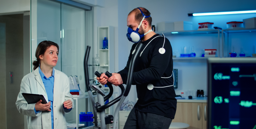
    Treadmill Test determines how your heart responds to stress and evaluates your cardiovascular status. You will be asked to walk on a treadmill until you reach a “target” heart rate based on your age. During the treadmill, blood pressure, heart rate, rhythm, and ECG changes will be continuously monitored.

    Indications for treadmill testing include:

    • Symptoms suggesting myocardial ischemia
    • Acute chest pain in patients excluded for acute coronary syndrome (ACS)
    • Recent ACS treated without coronary angiography or incomplete revascularization
    • Known CAD with worsening symptoms
    • Prior coronary revascularization (5 years or longer after coronary artery bypass grafting [CABG] or two years or less after percutaneous coronary intervention [PCI])
    • Prior coronary revascularization (5 years or longer after coronary artery bypass grafting [CABG] or two years or less after percutaneous coronary intervention [PCI])
    • Certain cardiac arrhythmias to assess chronotropic competenc
    • Newly diagnosed heart failure or cardiomyopath


    Venous Ablations

    Venous ablation is a treatment procedure to help regulate blood flow by heating or freezing certain veins. Venous ablation procedures are performed when other treatments, such as medication and exercise, don’t work to improve the condition of varicose veins. This procedure only treats the symptoms of varicose veins rather than curing them.

    Venous ablation works by using high-frequency sound waves to generate heat in the vein walls. The heat causes scar tissue formation which results in a permanent closure of that vein.Heat can also be applied with lasers or through an instrument inserted into a vein (endovenous thermal ablation). When cold is used, it freezes the surrounding tissue to kill the vein lining so blood no longer flows through it.Venous ablation is a low-risk procedure. The risks associated with the procedure are blood clots, infection, skin burns or scarring, and nerve damage. Venous ablation is not an effective treatment of leg swelling caused by heart failure (edema).
    Venous ablation can be conducted on several venous conditions. Procedures may include: – Laser therapy to close problem veins (percutaneous laser ablation) – Radiofrequency heat or cold applied to certain veins (radiofrequency ablation) or endovenous thermal ablation using either heat or a cold Venous ablation is an option for people who cannot take medications that improve vein problems or don’t want to undergo surgery due to risk factors. Venous ablation has been found to be successful in reducing symptoms and improving quality of life. Venous ablation is a procedure that uses heat or cold to close veins. It doesn’t cure varicose veins but instead treats their symptoms of them by closing off certain veins. Venous ablation also has low risks associated with it. A venous ablation is an option for people who cannot take medications that improve vein problems or don’t want to undergo surgery due to risk factors. Venous ablation has been found to be successful in reducing symptoms and improving quality of life

    Venous U/S

    Venous ultrasound (VUS) is a valuable tool for the assessment of lower limb venous disease. It can be used to both diagnose and guide treatment. Venous U/S has been valuable in assessing patients with chronic venous insufficiency (CVI) where clinical examination alone has led to conflicting results, especially in cases of superficial thrombophlebitis or isolated calf vein reflux. 

    Venous U/S enables the identification of incompetent valves, deep venous reflux and venous ectasia. Venography remains the gold standard method for evaluating the more proximal veins of the lower limb, but Venous U/S can be used as an alternative in selected cases, particularly when there are contraindications to venographic techniques.Venous U/S has been widely reported in the literature and has emerged as a valuable non-invasive tool for use in patients with venous disease. Venous ultrasound (VUS) is a valuable tool for assessing lower limb venous disease. VUS can be used both to diagnose and guide treatment of Venous Disease Venography remains the gold standard test, but Venous U/S can be used as an alternative when there are contraindications to Venography . Venous U/S has been widely reported and can serve as a non-invasive method for diagnosing lower limb veins. Venous U/S is a valuable tool for assessing the lower limb venous disease, and Venous U/S can be used to both diagnose and guide treatment. Venography remains the gold standard test for evaluating more proximal veins of the lower limb, but Venous U/S can be used as an alternative in selected cases when there are contraindications to Venography.


    Adenosine Myocardial Perfusion Imaging Test

    An Adenosine Myocardial Perfusion Imaging test uses a radioactive imaging agent (not a dye) to produce pictures of the heart muscle. The imaging agent gives off a small amount of radiation which can be seen with a special camera. The amount of radiation exposure during this test is very small and poses no health risk.

    The test will help your doctor determine if there are areas of your heart that do not receive enough blood supply due to coronary artery disease. Areas of your heart that may have been damaged from a previous heart attack may also be seen.

    Prior to the test, an IV will be started. During the rest portion of the test, you will receive an injection of the radioactive imaging agent, and pictures will be taken of your heart. For the stress portion of the test, a medication (Adenosine) will be given through the IV to simulate exercise by dilating the blood vessels of the heart. During the infusion, you will be continuously monitored for blood pressure, heart rate, rhythm, and ECG changes.

    You will be given an injection of the radioactive imaging agent during the infusion. Following the Adenosine infusion, pictures will be taken to determine blood flow to the heart muscle. The test will help your doctor determine if there are areas of your heart that do not receive enough blood supply due to coronary artery disease. Areas of your heart that may have been damaged from a previous heart attack may also be seen.

    Arterial Duplex Study

    An Arterial Duplex Study is also known as LEAI (lower extremity arterial imaging). This test will check the arterial circulation in the legs. This non-invasive test does not use needles, drugs, or dyes. The test will be performed by a highly-skilled ultrasound technologist.
    A small amount of gel is applied to the skin over the area to be studied. A handheld transducer is used to make the pictures. The pictures will show the walls of the arteries, blood flow through the arteries, and any plaque formation in the arteries. The velocity of the blood flow will be measured. This will pinpoint the level and severity of the disease if any.
    Your doctor may order additional testing if this test is abnormal. This test always includes an ABI (ankle-brachial index). Blood pressures are taken in both arms and ankles. The pressures are compared and calculations performed to estimate the severity of the disease.

    Carotid U/S

    Carotid ultrasonography (carotid U/S) is an ultrasound of the carotid arteries. Carotids are the arteries in your neck that supply blood to your brain. A small transducer, similar to the one used for listening with a stethoscope, is placed on the patient’s neck next to their windpipe (trachea). 

    When it’s pressed firmly into place, the transducer emits sound waves that bounce back and create echoes when they hit objects – in this case, different types of tissue in the carotid artery.Carotid ultrasounds are most often done in order to look at plaque build-up inside of blood vessels (“carotid intima-media thickness”), but they can also be done to look for atherosclerotic plaques, blood clots, and narrowing of the carotid arteries. Carotid ultrasounds are usually done on people who have risk factors for or a history of stroke or transient ischemic attack (TIA).
    Carotids ultrasound images may be interpreted by a sonographer and reported by an ultrasonographer or vascular technologist. Carotids U/S reports will often include measurements such as the thickness of the intima-media complex (IMT), which is used to assess plaque build-up. Carotids ultrasounds also provide information about any stenosis in the carotid artery, which can lead to decreased blood flow. Carotids U/S is also referred to as Carotids duplex U/S. Carotid ultrasounds are sometimes done in conjunction with carotid duplex Doppler ultrasounds, another type of ultrasound that provides information about blood flow in the arteries. Carotids duplex Doppler ultrasounds are most often used to look for carotid artery stenosis or occlusions. Ultrasound is a painless procedure that takes very little time.
    Carotid percussions can be performed at the bedside by compressing the neck over the artery and producing a thudding sound when it is released. This technique has been replaced by high-frequency ultrasound because it is more sensitive but less specific in detecting vascular abnormalities . Carotid auscultation has been shown to have a specificity of less than 50% for identifying vascular pathology. Carotids ultrasounds may be used to look at the following: Carotids arch – Carotids bifurcation – Carotid body – Carotids endarterectomy – Carotids “halo sign” – Carotid intima-media thickness (IMT) – Carotid lumen stenosis/obstruction – Carotids plaque build-up – Carotid plaques, calcified plaques, and complex composite plaques. Carotid Artery Ultrasound Images. Carotid artery ultrasound image showing noncompliant left carotid wall.

    Cardioversion

    A cardioversion is done to change or “convert” an irregular heartbeat to a normal regular rhythm. The Heart The heart is a muscular organ about the size of a closed fist. It pumps oxygen-rich blood to the body.A cardioversion is done to change or “convert” an irregular heartbeat to a normal regular rhythm. The Heart The heart is a muscular organ about the size of a closed fist.
    For the heart to do its work, it needs a “spark plug” or electrical impulse or signal from special tissue (sinus node) in the heart. The electrical impulse causes the heart muscle to contract. The hearts electrical system and muscle work together to effectively pump blood. Usually the heart beats regularly, but sometimes the heart beats irregularly. An irregular heart beat can occur after a heart attack or heart surgery. It may also occur with heart valve disease or other diseases not related to the heart. One cause of an irregular heart beat is atrial fibrillation. Some people with atrial fibrillation have no symptoms. Others may feel palpitations, shortness of breath, weakness or fatigue during episodes of atrial fibrillation. Your physician may first use medicines to convert your atrial fibrillation to a regular rhythm. If medicines do not work, an electrical cardioversion may be needed. This procedure uses small amounts of electrical current given through patches or paddles placed on the chest. The electrical current is used to restore your heart to a normal regular rhythm. When you are completely asleep, the physician will administer a very brief current of electricity. This electrical stimulation is intended to stop the irregular heartbeat and replace it with a normal heart rhythm. This is called converting the rhythm. The physician will administer the electrical current one to three times. Most people will convert to a normal rhythm with only one shock. If you do not convert after three attempts, the physician will not attempt any further electrical stimulation. The entire process lasts only a few minutes. The sedative medication will take from 10 minutes to two hours to wear off depending on the medication used and your individual response. The nurse will keep you in the room until you are completely awake and are able to sit up and talk. The nurse will make any follow-up appointments at this time and review any medication changes with you and your family members. On rare occasions the physician will administer special medications that assist in converting an irregular heart rhythm. If this occurs you may need to stay longer and be moved to another area that allows close monitoring of your heart rhythm for a few hours. This is a precaution that is used whenever this medication is administered.

    Cardiac Catherization

    A Cardiac Catheterization allows a physician to examine a patient’s heart for pumping function, check for blockage in the arteries bringing blood to the heart, and measure pressures within the heart. Patients are usually given a mild sedative prior to the procedure. This will help you feel more relaxed and maybe a bit sleepy.This test is done in a room with special cameras to take x-ray pictures of your heart. 

    The nurse or technician will cleanse and shave the area and cover you with sterile drapes. The doctor will then numb the area and insert a small hollow tube or sheath into the artery in your groin (or arm). The physician can then thread special catheters (long, thin plastic tubes) up to the heart.This test is done in a room with special cameras to take x-ray pictures of your heart. Most cardiac catheterizations are done through a large artery in the groin, However Dr. De is known for being one of the few who prefer the radial approach, done through the wrist.


    Through these specially shaped catheters, the physician can inject x-ray contrast dye and take x-ray pictures of the arteries that supply blood to the heart muscle and the main pumping chamber of the heart. Some people may notice a mild discomfort or “hot flash” following the dye injection which will last only a few seconds.


    During the catheterization, the physician can view the x-ray images on special TV screens. The entire procedure can take as little as 20 minutes or sometimes more than an hour, depending on how many and what type of measurements are taken. After the physician has completed the procedure, the sheaths are removed from the groin (or arm) and a nurse or cath lab technician will hold pressure on the site to allow the small hole in the artery to close. A special dressing, pressure device or sandbag may be placed on the site at this time.


    You will be asked to lie flat and still for several hours after the procedure to allow the hole in the artery to seal. Some physicians use a type of vascular closure device to seal the puncture in the artery. You should follow your physician’s specific instructions about activity following the use of one of these devices.

    Most patients will go home several hours after the procedure, but because you have been given medication to relax you, please plan to have someone drive you home.
    Arterial Duplex Study

    An Arterial Duplex Study is also known as LEAI (lower extremity arterial imaging). This test will check the arterial circulation in the legs. This non-invasive test does not use needles, drugs, or dyes. The test will be performed by a highly-skilled ultrasound technologist.
    A small amount of gel is applied to the skin over the area to be studied. A handheld transducer is used to make the pictures. The pictures will show the walls of the arteries, blood flow through the arteries, and any plaque formation in the arteries. The velocity of the blood flow will be measured. This will pinpoint the level and severity of the disease if any.
    Your doctor may order additional testing if this test is abnormal. This test always includes an ABI (ankle-brachial index). Blood pressures are taken in both arms and ankles. The pressures are compared and calculations performed to estimate the severity of the disease.

    Implantable Cardiac Monitor (Loop Recorder)

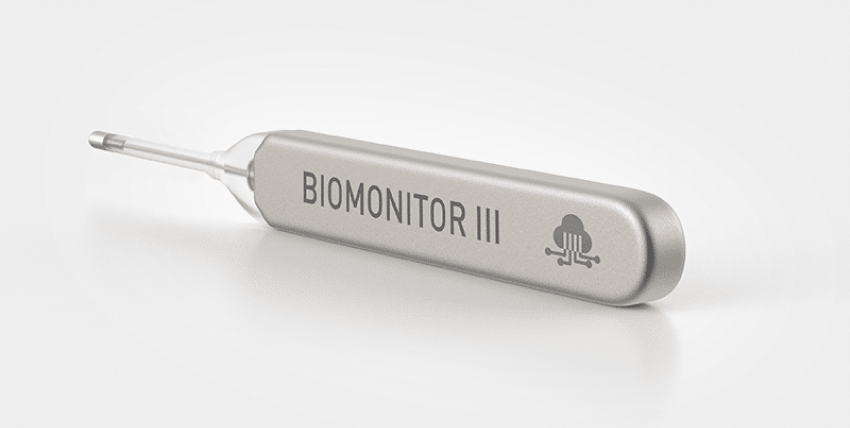
    Your cardiologist may recommend surgically implanting a wireless cardiac monitor called a loop recorder, which continuously records your heart’s rhythm for up to three years. Smaller than the size of an AAA battery, this device is surgically inserted beneath the skin of the upper chest to record the heart’s electrical activity, much like an electrocardiogram, or EKG.

    People who have had unexplained fainting spells or heart palpitations that can’t be detected by short term heart rhythm recording devices—such as Holter monitors—may be candidates for this device. Doctors may also recommend an implantable loop recorder for people with atrial fibrillation, which causes a rapid and irregular heartbeat. In addition, this device is used in people who have had a stroke for which a cause has not been determined.


    Your doctor makes a small incision in the upper chest and inserts the device just under the skin. This is a brief, outpatient procedure done in our office, you can return to your usual activities the next day. Your doctor may limit your activity until the incision heals.


    Cardiac loop recorders typically monitor the heart’s electrical activity for up to 36 months before replacement is required. The device is removed through the original incision site through a short surgery that is similar to the implantation procedure.
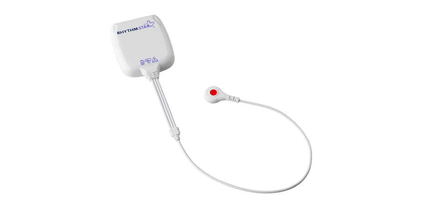
    Mobile Cardiac Telemetry

    Mobile Cardiac Telemetry is when a monitor is placed on a patient to transmit the heartbeat patterns remotely and in real-time. Mobile Cardiac Telemetry (MCT) is the cutting-edge of cardiac monitoring. MCT devices are small portable monitors that send a constant feed of data showing the ECG (Electrocardiography) readout of the patient in real-time.
    Electrocardiography is the process of recording the electrical activity of the heart over a period of time using electrodes placed over the skin. These electrodes detect the tiny electrical changes on the skin that arise from the heart muscle’s electrophysiologic pattern of depolarizing and repolarizing during each heartbeat.

    Peripheral Vascular Interventions

    What is Peripheral Vascular Intervention?

    Peripheral vascular intervention is a minimally invasive outpatient (same day) procedure used to treat peripheral artery disease, which causes plaque buildup in the arteries leading to the intestines, head, arms, and most commonly the legs. Plaque buildup is also called atherosclerosis.
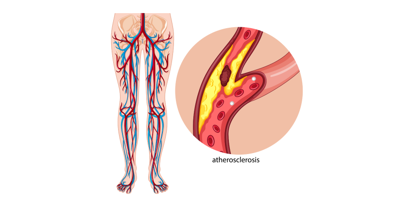
    The goal of peripheral vascular intervention is to restore the flow of blood to your lower extremities, eliminating pain, numbness, or need for amputation. Our specialists typically employ two different methods – angioplasty and atherectomy.


    Angioplasty & Stents
    Angioplasty is a minimally invasive intervention method that uses a balloon-tipped catheter to dilate your artery. Once inserted, the balloon is inflated at the blockage site to compress the plaque against the wall of the artery, increasing the flow of blood. In order to prevent the artery from becoming blocked again long-term, a small tube called a stent is sometimes put in place.


    Atherectomy
    Atherectomy is a non-surgical intervention that removes plaque using a catheter with either a sharp blade or a small drill on its tip. The plaque is either captured by the end of a catheter or is broken up and safely released into the bloodstream.
    Positron Emission Tomography (PET)

    What is a cardiac PET scan?

    A PET scan of the heart is a noninvasive nuclear imaging test. It uses radioactive tracers (called radionuclides) to produce pictures of your heart. Doctors use cardiac PET scans to diagnose coronary artery disease (CAD) and damage due to a heart attack. PET scans can show healthy and damaged heart muscle. Doctors also use PET scans to help find out if you will benefit from a percutaneous coronary intervention (PCI) such as angioplasty and stenting, coronary artery bypass surgery (CABG) or another procedure.

    Quick Facts

  • PET scans use radioactive material called tracers. Tracers mix with your blood and are taken up by your heart muscle.
  • A special “gamma” detector that circles the chest picks up signals from the tracer. A computer converts the signals into pictures of your heart at work.
  • A PET scan shows if your heart is getting enough blood or if blood flow is reduced because of narrowed arteries. It also shows dead cells (scars) from a prior heart attack.
  • A PET scan can help in determining if you’ll benefit from a cardiac procedure (PCI) or surgery to restore blood flow. The tracers used for PET scans can help identify injured but still living (viable) heart muscle that might be saved if blood flow is restored.
  • Why do people have cardiac PET scans?

    A PET scan is a very accurate way to diagnose coronary artery disease and detect areas of low blood flow in the heart. PET can also identify dead tissue and injured tissue that’s still living and functioning. If the tissue is viable, you may benefit from a PCI or coronary artery bypass surgery.

    How does a PET scan work?

    A radioactive tracer is injected into your bloodstream. The tracers used for PET are mostly natural body compounds such as glucose, water or ammonia, which are labeled or “tagged” with a small amount of radioactive material. Inside your body the radioactive tracer produces a type of energy called a gamma ray. Gamma rays are detected by a gamma detector and are used to produce a series of clear images of your heart. Images of thin slices made all the way through the heart can be produced from all different directions and angles.
    Computer graphics can be used to create a 3-dimensional image of your heart from the thin-slice images. Your doctor will be able to tell if your heart muscle is functioning or (if the heart muscle isn’t working) if your heart muscle is still alive by how well it takes up and uses the different tracers. The doctor will determine this by examining the images to find where the tracer is. Viable heart tissue will take in more of the tracer than tissue that’s no longer viable. Different colors or degrees of brightness on the PET scan show different levels of tissue function.

    What are the risks of cardiac PET?

    Cardiac PET is safe for most people. The amount of radiation is small, and your body will get rid of it through your kidneys within about 24 hours. If you’re pregnant or think you might be pregnant, or if you’re a nursing mother, tell your doctor before you have this test. It could harm your baby.

    How do I prepare for cardiac PET?

    • Tell your doctor about any medicines you take, including over-the-counter medicines, herbs and vitamins.
    • Your doctor may ask you not to take them before the test. Don’t stop taking your medicine until your doctor tells you to.
    • If you have diabetes and take insulin, talk with your doctor about how much insulin you should take before the scan and what you should eat. Your blood sugar levels will be monitored during the test. Test results are not always accurate in people with diabetes.
    • Your doctor may also ask you to avoid certain foods and drinks, such as caffeine-containing drinks or alcohol, for 24 hours before your test. Don’t eat, and drink only water for 4 to 6 hours before your test.
    • Wear comfortable, loose-fitting clothing, and don’t wear jewelry or other metal objects.

    What happens during cardiac PET?

    A doctor and a nuclear medicine technologist usually perform the scan in a hospital or at a PET center using special equipment.
    • The technologist will place small metal disks (electrodes) on your chest, arms and legs. The disks have wires that hook to a machine that records your electrocardiogram (ECG or EKG). The ECG keeps track of your heartbeat during the test and can signal the computer when to take a scan.
    • The technologist will put an intravenous line (IV) in your arm. The tracer will be injected through the IV line.
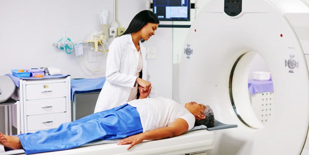
    • You’ll lie on a flat table that’s connected to the PET scanner and a computer. The table will slide into the scanner, which is shaped like a giant doughnut.
    • Within the PET scanner, detectors record the radioactive patterns of the tracer in your heart. The information is transformed into images on a computer screen. Several scans are done over time to provide pictures of thin slices of your entire heart from all angles. It’s very important to hold completely still with your arms above your head while each scan is being done.
    • Doctors will take a baseline picture of your heart before the tracer is injected. This takes about 15 to 30 minutes.
    • Doctors will take a baseline picture of your heart before the tracer is injected. This takes about 15 to 30 minutes.
    • Next, the tracer will be injected and your heart will be scanned again.
    • If you will have a nuclear chemical stress test (also called a pharmacologic stress test), you’ll get a medicine that increases the blood flow in your heart, similar to what happens during exercise. These medicines may include adenosine, dipyridamole (Persantine) or dobutamine. The doctor will examine how well your heart takes up the tracer before and after receiving the medicine. If you have severe coronary artery disease, some areas of your heart may not get enough blood during a stress so the tracer won’t show up in those areas.
    • The test takes between 1 and 3 hours.

    What happens after my PET scan?

    • You can usually go back to your normal activities right away.
    • Drink plenty of water for the next 24 hours to flush the radioactive material from your body.
    • Make an appointment with your doctor to discuss the results of the test and next steps.

    Transesophageal Echocardiograms

    A TEE is a test which uses a flexible tube that produces ultrasound waves to create pictures of the heart from inside of the esophagus and stomach. Sound waves are transmitted from an instrument, called a transducer probe, into your body.Sound waves are transmitted from an instrument, called a transducer probe, into your body.

    The sound waves reflect (echo) off the tissues and organs to create pictures which can be seen on a screen. The TEE provides very clear pictures of the heart structures and blood flow. The pictures are usually clearer than those obtained from a standard echocardiogram, which is performed from the chest wall. This test is often used to view hard-to-see structures or to obtain more detailed pictures of the heart and aorta.


    Common reasons for the test include measuring the size and pumping strength of the heart and looking at the shape and motion of the heart valves. This test may also be used to look for fluid around the heart and blood clots or masses inside of the heart or aorta.A blood pressure cuff will be placed on your arm. Small sticky patches, called electrodes, will be placed on your chest. An oxygen transducer will be placed on your finger to measure the amount of oxygen in your body. Your heart rhythm, blood pressure, oxygen levels, and breathing will be monitored during the test. You will be given oxygen.


    An intravenous line (or IV) will be placed into a vein in your arm. A local anesthetic will be sprayed to numb the back of your throat. Medicine will be given through your IV to help you relax and feel sleepy. The doctor will gently insert the probe into your mouth. As you swallow, the probe will be directed into your esophagus. You may feel the probe moving, but it is generally not painful. When the probe is positioned in the esophagus just behind the heart, pictures of the heart will be taken. The test will take about 1 1/2 hours.

    Treadmill Test

    Treadmill stress testing is indicated for the diagnosis and prognosis of coronary artery disease (CAD). This is the initial investigation of choice in patients with a normal or near-normal resting ECG capable of performing adequate exercise.
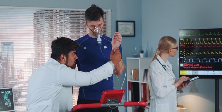
    Treadmill Test determines how your heart responds to stress and evaluates your cardiovascular status. You will be asked to walk on a treadmill until you reach a “target” heart rate based on your age. During the treadmill, blood pressure, heart rate, rhythm, and ECG changes will be continuously monitored.

    Indications for treadmill testing include:

    • Symptoms suggesting myocardial ischemia
    • Acute chest pain in patients excluded for acute coronary syndrome (ACS)
    • Recent ACS treated without coronary angiography or incomplete revascularization
    • Known CAD with worsening symptoms
    • Prior coronary revascularization (5 years or longer after coronary artery bypass grafting [CABG] or two years or less after percutaneous coronary intervention [PCI])
    • Prior coronary revascularization (5 years or longer after coronary artery bypass grafting [CABG] or two years or less after percutaneous coronary intervention [PCI])
    • Certain cardiac arrhythmias to assess chronotropic competenc
    • Newly diagnosed heart failure or cardiomyopath


    Venous U/S

    Venous ultrasound (VUS) is a valuable tool for the assessment of lower limb venous disease. It can be used to both diagnose and guide treatment. Venous U/S has been valuable in assessing patients with chronic venous insufficiency (CVI) where clinical examination alone has led to conflicting results, especially in cases of superficial thrombophlebitis or isolated calf vein reflux. 

    Venous U/S enables the identification of incompetent valves, deep venous reflux and venous ectasia. Venography remains the gold standard method for evaluating the more proximal veins of the lower limb, but Venous U/S can be used as an alternative in selected cases, particularly when there are contraindications to venographic techniques.Venous U/S has been widely reported in the literature and has emerged as a valuable non-invasive tool for use in patients with venous disease. Venous ultrasound (VUS) is a valuable tool for assessing lower limb venous disease. VUS can be used both to diagnose and guide treatment of Venous Disease Venography remains the gold standard test, but Venous U/S can be used as an alternative when there are contraindications to Venography . Venous U/S has been widely reported and can serve as a non-invasive method for diagnosing lower limb veins. Venous U/S is a valuable tool for assessing the lower limb venous disease, and Venous U/S can be used to both diagnose and guide treatment. Venography remains the gold standard test for evaluating more proximal veins of the lower limb, but Venous U/S can be used as an alternative in selected cases when there are contraindications to Venography.


    Venous Ablations

    Venous ablation is a treatment procedure to help regulate blood flow by heating or freezing certain veins. Venous ablation procedures are performed when other treatments, such as medication and exercise, don’t work to improve the condition of varicose veins. This procedure only treats the symptoms of varicose veins rather than curing them.
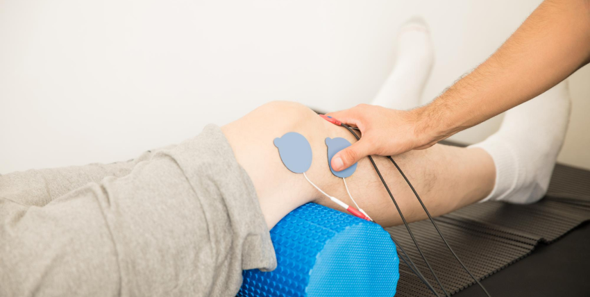
    Venous ablation works by using high-frequency sound waves to generate heat in the vein walls. The heat causes scar tissue formation which results in a permanent closure of that vein.Heat can also be applied with lasers or through an instrument inserted into a vein (endovenous thermal ablation). When cold is used, it freezes the surrounding tissue to kill the vein lining so blood no longer flows through it.Venous ablation is a low-risk procedure. The risks associated with the procedure are blood clots, infection, skin burns or scarring, and nerve damage. Venous ablation is not an effective treatment of leg swelling caused by heart failure (edema).
    Venous ablation can be conducted on several venous conditions. Procedures may include: – Laser therapy to close problem veins (percutaneous laser ablation) – Radiofrequency heat or cold applied to certain veins (radiofrequency ablation) or endovenous thermal ablation using either heat or a cold Venous ablation is an option for people who cannot take medications that improve vein problems or don’t want to undergo surgery due to risk factors. Venous ablation has been found to be successful in reducing symptoms and improving quality of life. Venous ablation is a procedure that uses heat or cold to close veins. It doesn’t cure varicose veins but instead treats their symptoms of them by closing off certain veins. Venous ablation also has low risks associated with it. A venous ablation is an option for people who cannot take medications that improve vein problems or don’t want to undergo surgery due to risk factors. Venous ablation has been found to be successful in reducing symptoms and improving quality of life

    Need a Doctor for Check-up?

    Our cardiologists will help you solve cardiology related problems painless and carefully.

    What Should I Do?

    1.
    Register for the First Time
    2.
    Choose Date & Time
    3.
    Book an Appointment
    4.
    You’re Ready for Check-up.